-
The University
- Welcome
- Who we are
- Media & PR
- Studying
-
Research
- Profile
- Infrastructure
- Cooperations
- Services
-
Career
- Med Uni Graz as an Employer
- Educational Opportunities
- Work Environment
- Job openings
-
Diagnostics
- Patients
- Referring physicians
-
Health Topics
- Health Infrastructure
Clinical Forensic Medicine
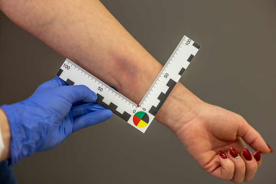
In the area of clinical forensic medicine, the following projects of different issues are currently being conducted.
PILOT PROJECT ON TELEMEDICINE IN CLINICAL AND FORENSIC EXAMINATIONS
In this project, funded by the province of Styria, a telemedicine system called Forensic PETRA is being developed to enable forensic investigators to conduct clinical and forensic examinations live through the eyes of the investigator.

PILOT PROJECT MODEL REGION SOUTH
In this project funded by the federal government, the Clinical Forensic Medical Examination Center at the D & F Institute for Forensic Medicine is being expanded and will begin offering clinically forensic examinations that can be used in court, in accordance with the latest scientific standards, across the board.


